Imaging and Interpretation for You and Your Physician
Minneapolis Radiology has combined our imaging experience with cutting edge technology to deliver the highest level of diagnostic interpretation for referring physicians and their patients. For each diagnostic service our radiologist will send your health care provider an accurate, timely interpretation. You and your provider can then discuss the results to best guide your treatment.
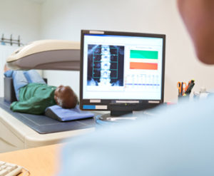
What is Bone Density (DEXA)? Bone densitometry, also called DEXA (dual-energy X-ray absorptiometry), is a medical imaging exam that uses an enhanced form of X-ray to measure bone density and bone loss. What should I bring to my appointment? Your insurance card and a valid photo ID. Please arrive 15 minutes early to complete...
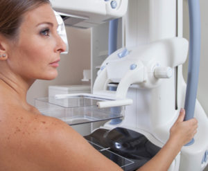
Learn MoreMammograms are examinations of the breasts using low-dose X-rays. Most experts agree that mammography is the most effective tool for early breast cancer detection. At Minneapolis Radiology, we use state-of-the-art computer-aided detection (CAD) technology to assist in diagnosis. Current guidelines recommend baseline mammograms beginning at age 40. Women of all ages, particularly those with a...
Learn MoreWhat Is a Cardiac CT Calcium Score? A cardiac CT calcium score, also known as a coronary calcium scan, is a quick, convenient, and noninvasive way of evaluating the amount of calcified (hard) plaque in your heart vessels. The level of calcium correlates to the extent of plaque build-up in your arteries, which can cause...
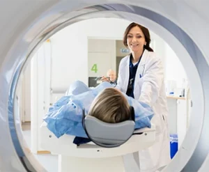
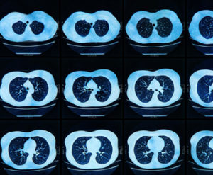
Learn MoreWhat is a CT? Computed Tomography (CT), also called a CAT scan, and is a medical imaging exam that takes computer assisted x-rays that produces two and three dimensional detailed images of the soft tissues and bones within the body. What should I bring to my appointment? Your insurance card and a valid photo...
Learn MoreWhat is a CTA? CTA stands for Computed Tomography Angiography. It is a specialized CT exam that obtains enhanced images of the major vessels in your body called arteries. The vessels most frequently imaged in this manner are of the brain, neck (carotid), aorta, kidney (renal) and extremities. How should I prepare for my...
Learn MoreWhat is a CT Lung Cancer Screening? A chest CT lung cancer screening is a low-dose (low-radiation) chest CT exam used to detect early stage lung cancer before signs or symptoms appear. The screening may detect lung abnormalities or nodules that would not otherwise be visible on a plain chest x-ray. Many nodules will be non-cancerous and...
Learn MoreWhat is Hysterosalpingography (HSG)? Hysterosalpingography, also called uterosalpingography, is an x-ray examination of a woman's uterus and fallopian tubes that uses a special form of x-ray called fluoroscopy and a contrast material. An x-ray (radiograph) is a noninvasive medical test that helps physicians diagnose and treat medical conditions. Imaging with x-rays involves exposing a part...
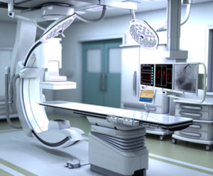
Learn MoreInterventional Radiology (IR) is a subspecialty field encompassing minimally-invasive procedures performed with image guidance. Our fellowship-trained IR doctors are experts in a comprehensive number of diagnostic and therapeutic procedures, including: Angiography Angioplasty & Stenting Biliary Drainage and Stenting Cancer Treatment (tumor ablation and chemoembolization) Cervical and Lumbar Spine Epidural Steroid Injections (ESI) Chest Tube Placement...
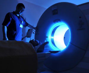
Learn MoreWhat is an MRI? Magnetic Resonance Imaging (MRI) is a medical imaging exam that uses a strong magnetic field and radio waves to collect detailed information about the organs and tissues of the body. MRI does not use x-ray or radiation. This information is displayed on a specialized computer system that is then studied and analyzed by a...
Learn MoreWhat is an MRA? MRA (Magnetic Resonance Angiography) is a specialized type of MRI exam used to obtain images of the arteries of the brain, carotid arteries, aorta, renal arteries and vessels of the extremities. MRA is used to detect problems with blood vessels that may be causing reduced blood flow. How should I...
Learn MoreNuclear Medicine imaging uses small amounts of radioactive material to diagnose and detect a full spectrum of diseases. The material is typically injected or swallowed and images are then acquired to aid in the diagnosis of your condition.
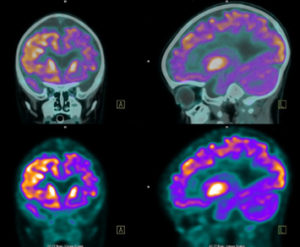
Learn MorePET imaging is now combined with CT for better anatomic localization. PET/CT is used most frequently to diagnose certain cancers, determine spread of disease and check response to therapy. These studies can also been helpful to diagnose heart disease and brain disorders. PET/CT involves injection of a small amount of radioactive material prior to scanning...
Learn MoreWhat is a pain management procedure? A pain management procedure, performed by a radiologist, is an image-guided injection of medication through a precisely placed, small needle. These procedures can provide pain relief, with the goal of returning you to normal activity. They can also give additional knowledge about your condition and finding the source of...
Learn MoreWhat is an Ultrasound? Ultrasound (also called a sonogram) is a medical imaging exam that uses sound waves to create images. These images show the structure and movement of the body’s organs and blood flow. Abdomen Aorta Arterial Bladder Carotid Extremities Newborn Hips Pelvis Pelvis/TV Renal OB/GYN Testicular Venous ABI What should I bring...
Learn MoreCompression Stockings, Conservative Vein Therapy, Interventional Radiology and Vascular Consultations, Phlebectomy, Pain Management (spine and joint injections), Sclerotherapy, Ultrasound Guided Sclerotherapy, Vascular Ultrasound, Veinwave, Venous Ablation, Venous Leg Ulcers. For more information, visit: Minneapolis Vein Center
Learn MoreWhat is an x-ray? An X-ray is a non-invasive exam that provides valuable information to help physicians diagnose and treat medical conditions. Imaging with x-rays involves using a small dose of radiation to produce images of the inside of the body. X-rays are the oldest and most frequently used form of medical imaging. What should...
Learn More















 Copyright © 2025 · Minneapolis Radiology Associates ·